


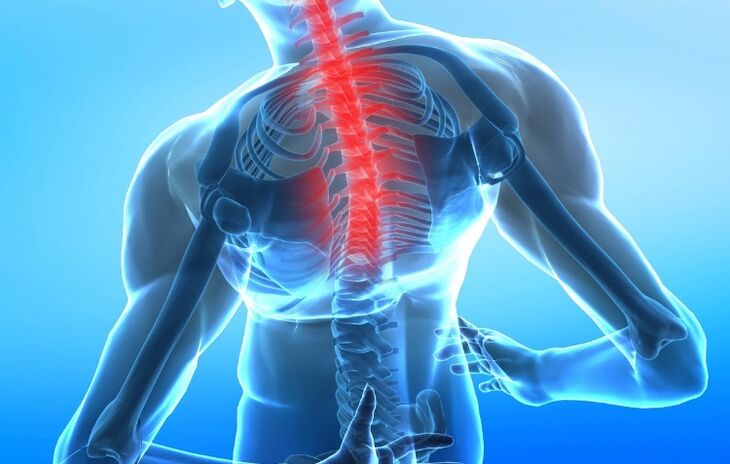
Osteochondrosis of the spine is a complex of dystrophic and degenerative changes in the intervertebral discs and adjacent surfaces of the vertebral body associated with tissue destruction and disruption of their structure. Depending on the degree of damage, there are cervical, thoracic and lumbar osteochondrosis.
Symptoms
The main signs by which the presence of osteochondrosis of the cervical spine can be assumed is a local change in the configuration of one of the segments of the spine (development of lordosis, kyphosis or scoliosis) - a clear visual curvature of the spine. in the longitudinal or transverse plane. The second most common symptom is pain syndrome, which can be localized not only in the vertebral region, but also on parts of the body innervated by the corresponding nerve root. Another complaint of these patients is a feeling of discomfort and a feeling of fatigue in the neck.
In cervical osteochondrosis, the pain usually manifests itself in the neck area and can be given in the shoulder and shoulder blade, it can be confused with the pain in myocardial infarction, because it has similar symptoms. Also, cervical osteochondrosis may be accompanied by frequent headaches, dizziness. When the arteries that supply the brain are compressed, signs of brain damage (neurological symptoms) can appear: fainting, nausea, tinnitus, mood swings, anxiety, and more.
According to the intensity of pain, they are divided into 3 degrees:
- Pain occurs only with pronounced movements in the spine;
- The pain is relieved by a certain position of the spine;
- The pain is permanent.
Forms
Depending on the syndromes caused by osteochondrosis, there are:
- Compression syndromes - occur with compression (radiculopathy - compression of nerve roots, myelopathy - compression of muscles, neurovascular - compression of blood vessels and nerves);
- Reflex (muscular-tonic, neurodystrophic, neurovascular);
- Myoadaptive syndrome (overexertion of healthy muscles when they take over the functions of affected muscles).
Causes
The mechanism of disease development is damage to the intervertebral disc for various reasons and its displacement with loss of depressive (relieving pressures) functions of the spine. The immediate cause of disc damage can be age-related degenerative changes associated with impaired blood supply to the intervertebral discs, mechanical damage from injury, and strenuous physical exertion on the spine — for example, being overweight.
An important role in the development of osteochondrosis is played by a sedentary lifestyle, in which a violation of the blood supply and the functioning of the intervertebral joints develops. The mechanism of disease development is as follows: if the fibrous ring connecting the vertebral bodies is damaged, the intervertebral disc is pushed back and forth - into the lumen of the spinal canal, or laterally - with the formation of the middle and lateral disc. hernia. The disc can be pushed into the body of the vertebra itself with the formation of Schmorl's hernia - microscopic fractures of the cartilaginous tissue of the intervertebral disc into the spongy tissue of the spine. In the case of posterior displacement of the disc, compression of the spinal cord and roots extending from it is possible, with the development of a typical pain syndrome.
Diagnosis
The diagnosis of osteochondrosis of the spine is made on the basis of complaints, anamnesis data, clinical examination and instrumental examination methods. Diagnostic measures are to discover the reasons that led to the development of neurological symptoms.
The anamnesis can determine the presence of injury, the nature of the work - constant physical overload (lifting weights), poor posture, peculiarities of work and the position of the spine at the table and when walking, the presence of infections.
General clinical studies (clinical blood test, general urine analysis), biochemical blood test have no independent value. They are prescribed to assess the current condition, diagnose the underlying disease and the resulting complications.
The diagnosis is based on the clinical picture of the disease and is carried out by the method of sequential exclusion of diseases similar to clinical signs. Of the instrumental diagnostic methods, X-ray examination is the most common and available (spondylography is a non-contrast study). It reflects the narrowing of the intervertebral joint spaces and allows you to identify osteophytes (bone growths) on the vertebral bodies, but gives only indirect information about the degree of damage to the intervertebral discs.
Accurate diagnosis can be made by CT and MRI (computerized and magnetic resonance imaging) diagnostics, even in the early stages of the disease. CT allows you to determine minimal anomalies in bone and cartilage tissue, MRI - to perform a detailed processing of soft tissue structures and determine the location of disc herniation.
Duplex ultrasound scan of the cerebral arteries is performed if there is a suspicion of a violation of blood flow to the brain.
Differential diagnosis is made with diseases that have similar clinical manifestations: pathologies that take place with pain that spreads in the area of the shoulders and shoulders (diseases of the liver, gallbladder, pancreatitis - inflammation of the pancreas); cervical lymphadenitis - enlargement of cervical lymph nodes, rheumatoid arthritis; oncological diseases (tumors of the vertebrae, roots, spinal cord and membranes), tumors of the pharynx and pharyngeal space, Pancost cancer (compression of the brachial plexus in cancer of the upper lobe of the lung), metastases in the cervical region; tuberculous spondylitis - an inflammatory disease of the spine caused by the mycobacterium tuberculosis; arachnoid cysts; pseudocysts dura mater; spinal anomalies; fibromyalgia is a disease that causes pain in muscles, ligaments and tendons, chest compression syndrome - a disorder caused by excessive pressure on the neurovascular bundle that passes between the anterior and middle scapular muscle, over the first rib and below the collarbone, myofascial neck syndrome and shoulder girdle -a chronic, pathological condition caused by the formation of local muscle cramps or seals, represented by painful points.
Main laboratory tests used:
- Clinical blood test;
- Blood chemistry.
Main instrumental studies used:
- X-ray of the spine (spondylography);
- Magnetic resonance imaging (MRI);
- Computed tomography (CT);
- Ultrasound duplex scanning of brain arteries (if a violation of blood flow to the brain is suspected).
Additional instrumental studies were used:
- Densitometry - measurement of bone density (according to indications).
Treatment
Treatment of osteochondrosis of the spine depends entirely on the stage and stage of development of osteochondrosis. In the initial phase, it is possible to use preventive measures, physiotherapy exercises, exercises on simulators, fitness. With severe pain, the patient needs physical rest. Anti-inflammatory and antispasmodic drugs are prescribed. It is possible to perform paravertebral blockades with anesthetics in order to open the pathological circle, when the pain causes muscle spasm, while the intervertebral disc is compressed more, which in turn intensifies the pain itself.
Warming ointments are applied topically to the skin in the area of the spine to improve the local blood supply and reduce tissue edema. These patients are shown wearing a corset. In patients with the initial stage of osteochondrosis, chondroprotectors are effective - drugs that improve the regeneration of cartilage tissue, as well as drugs that improve the local blood supply, venotonics, B vitamins. In cases where the pain syndrome does not stop medically for a long timebrain with intervertebral hernia, surgical removal of the damaged intervertebral disc is shown. In cases of total compression of the spinal cord with a disc, early surgery is indicated.
You should not wait for a person to start urinating spontaneously or defecating - in which case the damage to the spinal cord may already be irreversible. Magnetotherapy, ultrasound, massage, manual therapy, acupuncture and physiotherapy exercises are prescribed as physiotherapeutic procedures.
Complications
Possible vegetative-vascular dystonia and cardiac dysfunction, cerebrovascular infarction, hypotension and hypertension (decrease and increase in blood pressure), vestibular disorders (impaired coordination of movements), vertebral artery syndrome (disease caused by narrowing of the vertebral artery), periarthrosisshoulder joint.
Prophylaxis
To prevent osteochondrosis, it is necessary to address the factors that cause it, which are: avoiding spinal injuries, stress on the spine (lifting weights), and the fight against excess weight. For people who are already suffering from the initial phase of osteochondrosis, it is recommended to wear a corset at home and during physical exertion. In order for the spine to rest during sleep, it is recommended to sleep on orthopedic mattresses and pillows.
What questions should you ask your doctor
Are there any exercises that help alleviate the symptoms?
What medications will help address osteochondrosis of the cervical spine?
What will happen if you do not start treating the disease in time?
Patient tips
Exercise, weight loss in the presence of excess weight, the use of cold or warm compresses help alleviate the symptoms of osteochondrosis of the thoracic spine. It is also important to eat properly, monitor the spine, treat chronic conditions and avoid injuries.